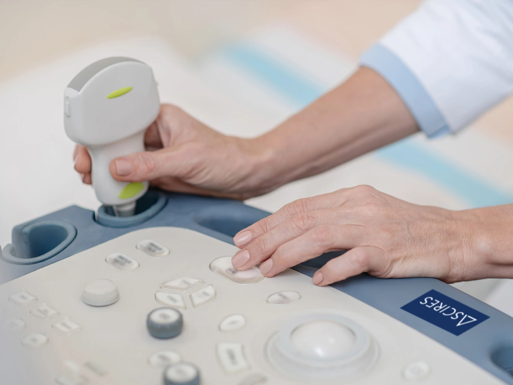
At Ascires Hospital in Valencia, we have advanced high-resolution technology to provide precise and accurate diagnoses using ultrasound.
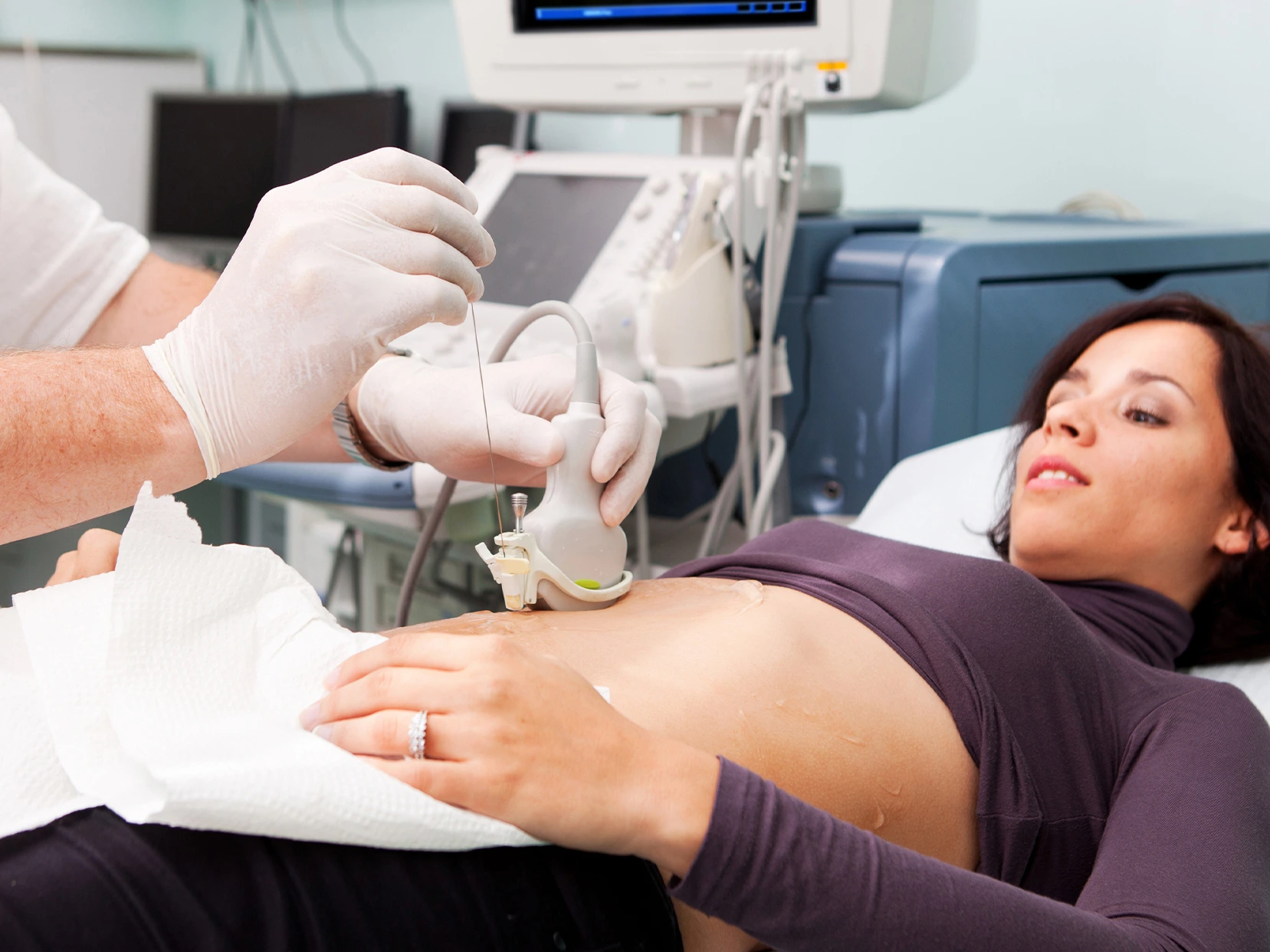
Ultrasound is a diagnostic imaging test that, without using radiation, employs sound waves to observe internal organs in detail. While it is well known in the context of pregnancy, where it is particularly useful for visualising the foetus, this technique is also ideal for examining organs such as the kidneys, liver, muscles or heart.
Features
What makes our ultrasound tests stand out
More information
At the Radiology Service of Ascires in Valencia, we perform different types of ultrasound examinations:
Related services
| Name | Address | Phone | |
|---|---|---|---|
| Castelló | |||
| Ascires Castelló | Dirección: Carrer de Betxí, 7, 12004 Castelló de la Plana, Castelló | Teléfono: 961 86 89 98 | Email: moc.sericsa@olletsac |
| Valencia | |||
| Ascires Campanar | Dirección: Radiodiagnóstico: Marqués de San Juan, 6 y 4 Consultas: Avenida Campanar 116 València | Teléfono: 96 340 40 77 | Email: moc.sericsa@ranapmac |
| Ascires Gandia | Dirección: Av. d’Alacant, 123 – Nave 6 46702 – Gandia | Teléfono: 96 186 89 99 | Email: moc.sericsa@aidnag |
| Ascires Sagunto | Dirección: C/ Advocat Fausto Caruana, 11 46520 – Puerto de Sagunto | Teléfono: 96 186 89 91 | Email: moc.sericsa@tnugas |
| Ascires Universitats | Dirección: C/ Menéndez y Pelayo, 25. 46010 Valencia | Teléfono: 963 40 47 90 | Email: moc.sericsa@ytisrevinu |
| Ascires Xàtiva | Dirección: Ronda Séquia de la Vila, s/n, 46800 – Xàtiva | Teléfono: 96 340 43 44 | Email: moc.sericsa@avitax |
An ultrasound is a diagnostic imaging test that uses high-frequency sound waves to visualise internal organs, soft tissues and structures such as the heart, liver, kidneys, uterus or the foetus during pregnancy in real time. Unlike other imaging tests, ultrasound does not use radiation, making it completely safe and non-invasive.
At our private hospital in Valencia, we perform ultrasounds for both diagnostic purposes and the monitoring of chronic conditions, gynaecological, digestive and musculoskeletal checks, and vascular evaluations. It is a quick, painless technique, highly useful in outpatient and clinical settings.
During pregnancy, several key ultrasounds are performed to assess the baby’s development and the mother’s health. The most common are three scans, one in each trimester. The first-trimester ultrasound (between weeks 11 and 14) confirms the pregnancy, determines gestational age, and evaluates the risk of chromosomal abnormalities through nuchal translucency measurement.
The second-trimester morphological ultrasound (around week 20) is one of the most important: it allows detailed study of fetal anatomy, detection of possible malformations, and assessment of the proper development of organs such as the brain, heart, spine and kidneys. Finally, in the third trimester, a control ultrasound is usually performed to check fetal growth, the baby’s position, and the amount of amniotic fluid.
Additionally, other complementary ultrasounds may be indicated depending on the circumstances: Doppler ultrasounds to study blood flow, transvaginal ultrasounds in early stages, or additional scans if there is any complication. At our hospital, we have professionals specialised in prenatal diagnosis and high-resolution equipment to ensure precise and safe monitoring of the pregnancy.
“Is any preparation required before an ultrasound?
For other examinations, such as muscle, thyroid, breast or testicular ultrasounds, no preparation is usually required.
In some cases, it is possible to have an ultrasound without a medical prescription, especially for preventive check-ups or voluntary examinations, such as abdominal, breast or gynaecological ultrasounds requested at the patient’s initiative. However, it is recommended to have a medical indication, as this helps guide the study and ensures its diagnostic usefulness.
At the hospital, we offer the option to perform certain ultrasounds without a prescription, but always under medical supervision. If the patient does not have a prescription, they can request a prior consultation with our specialists to assess their case and determine the most appropriate examination.
Conventional ultrasound provides real-time images of the body’s organs and tissues, while Doppler ultrasound also allows assessment of blood flow in arteries and veins. The latter is particularly useful for detecting circulatory problems such as thrombosis, varicose veins, arterial narrowing or venous insufficiency.
Both techniques use the same ultrasound principle, but Doppler incorporates additional analysis of blood movement, using colour coding or graphical representation. The doctor will determine which type of ultrasound is necessary based on the clinical suspicion or the patient’s symptoms.
Yes, ultrasounds are completely safe for children and babies, including newborns. They are widely used in paediatrics due to their non-invasive nature and the fact that they do not involve radiation exposure. For example, they are used to assess the abdomen, hips (hip ultrasound in neonates), the skull in infants, or for kidney and testicular studies.
The medical team adapts the technique to the patient’s age and, in the case of babies or young children, strives to create a calm and comfortable environment so that the test can be carried out without stress. In many cases, results are obtained immediately, allowing prompt intervention if necessary.
Ascires has the ENS certification with the MEDIA category for its medical centers in the Valencian Community and Madrid.